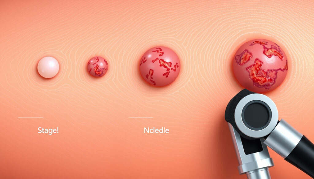
Basal Cell Carcinoma Stages Pictures: Stage-by-Stage Guide
When you notice a strange spot on your skin, it’s natural to wonder what it could be. For millions of people each year, that spot turns out to be basal cell carcinoma (BCC) — the most common form of skin cancer.
Annual U.S. cases: over 4 million ·
5-year survival rate (localized): 99% ·
Most common skin cancer: yes ·
Aggressive BCC subtypes: morpheaform, basosquamous, infiltrative ·
Metastasis rate: <0.1%
Quick snapshot
- BCC rarely metastasizes but aggressive subtypes have higher risk (American Cancer Society)
- Staging is based on tumor size, depth, and invasion (PMC / NIH)
- Visual identification is a screening tool, not a substitute for biopsy (American Academy of Dermatology)
- Exact staging thresholds may vary slightly between AJCC and Brigham guidelines (PMC / NIH)
- Long-term outcomes for untreated superficial BCC are not well documented (American Cancer Society)
- BCC typically grows slowly over months to years; aggressive subtypes may grow faster (StatPearls)
- If you suspect BCC, see a dermatologist for a biopsy (American Academy of Dermatology)
- Treatment options range from topical creams to Mohs surgery depending on stage (Memorial Sloan Kettering)
Five key facts about BCC, one pattern: the outlook is excellent when caught early, but aggressive subtypes require prompt attention.
| Most common cancer in the U.S. | Yes, over 4 million cases annually |
| 5-year survival (localized) | 99% |
| 5-year survival (metastatic) | ≈20% |
| Recurrence rate (clear margins) | <5% |
| High-risk locations | Face, ears, scalp, genitals |
What does stage 1 basal cell carcinoma look like?
Typical appearance – pearly or waxy bump
- Stage 1 BCC often appears as a small, raised, translucent or pearly bump with visible blood vessels. The American Cancer Society describes it as a “shiny, pearly bump” that may be pink, red, or flesh-colored (American Cancer Society).
- It can be mistaken for a pimple or scar that never heals (American Academy of Dermatology).
Color variations – pink, red, or flesh-colored
- Most BCCs are pink or red, but on darker skin tones they may appear brown, black, or blue (American Cancer Society).
- Pigmented BCC can look like a mole or melanoma, so biopsy is essential (StatPearls).
Early signs – small, translucent nodule
- Early BCC is usually less than 2 cm in diameter. The AJCC defines T1 as tumor ≤2 cm with fewer than 2 high-risk features (PMC / NIH).
- It may be flat or slightly raised, and some are described as “scar-like” (American Cancer Society).
Differences on darker skin tones
- On darker skin, BCC can be hypopigmented or hyperpigmented, often with a rolled border (American Academy of Dermatology).
- Delayed diagnosis is more common in people of color because the appearance is less typical (American Cancer Society).
Because stage 1 BCC is so small and innocuous, patients often delay consultation. The American Academy of Dermatology emphasizes that a biopsy is the only way to confirm the diagnosis.
The pattern: Stage 1 BCC is subtle and often dismissed. Early detection significantly improves treatment outcomes, but it requires knowing what to look for.
The catch: Even a small, pearly bump can be BCC, and a biopsy is the only way to be sure.
What does stage 3 basal cell carcinoma look like?
Larger size and deeper invasion
- Stage 3 BCC is defined as tumor of any size that has invaded the maxilla, mandible, orbit, or temporal bone, or has spread to one lymph node ≤3 cm (PMC / NIH).
- Visually, it may be a large, ulcerated nodule with rolled edges, often >2 cm (American Cancer Society).
Ulceration and crusting
- A non-healing sore that crusts, bleeds, or leaks fluid is a classic sign of advanced BCC (American Cancer Society).
- The center may be depressed, and the edges raised and pearly (American Academy of Dermatology).
Pigmented or non-pigmented variants
- Some stage 3 BCCs are heavily pigmented, mimicking melanoma (StatPearls).
- Others are flesh-colored with telangiectasias (visible blood vessels) (American Cancer Society).
Common locations and asymmetry
- High-risk locations include the central face, ears, scalp, and genitals (American Cancer Society).
- Asymmetry and irregular borders are more pronounced in advanced stages (American Academy of Dermatology).
Despite being advanced, stage 3 BCC still has a favorable prognosis if treated aggressively. The survival rate drops significantly only when metastasis occurs.
What this means: Stage 3 BCC is locally advanced and destructive. Prompt referral to a specialist is critical to prevent further damage.
What does aggressive basal cell carcinoma look like?
Morpheaform subtype – scar‑like plaque
- Morpheaform BCC appears as a flat, firm, pale or yellow scar-like area without distinct borders (American Cancer Society).
- It is more likely to recur after treatment (StatPearls).
Basosquamous carcinoma – mixed features
- Basosquamous carcinoma has features of both BCC and squamous cell carcinoma, making it more aggressive (StatPearls).
- It may look like a pearly nodule with a crusted surface (American Academy of Dermatology).
Infiltrative growth pattern
- Infiltrative BCC grows irregularly into the dermis, often with ill-defined borders (American Cancer Society).
- It is common in high-risk areas like the scalp and can be difficult to clear surgically (StatPearls).
Recurrence after previous treatment
- Aggressive BCC often recurs at the same site, especially if initial margins were not clear (American Cancer Society).
- Recurrence is a red flag for more aggressive histology (American Academy of Dermatology).
The trade-off: Aggressive BCC subtypes require more extensive surgery but are still curable. The key is identifying them early by their scar-like or infiltrative appearance.
How do I know what stage my basal cell carcinoma is?
Clinical examination and dermatoscopy
- A dermatologist will examine the lesion with a dermatoscope to look for BCC features like arborizing vessels and ulceration (American Academy of Dermatology).
Biopsy and histopathology
- Staging requires a biopsy to determine depth, size, and cell type. The pathologist will report the subtype and if there is perineural invasion (American Cancer Society).
Imaging for advanced cases
- Imaging (CT, MRI) is used only when invasion of bone or lymph nodes is suspected (AJR).
Staging systems (TNM vs. Brigham and Women’s)
- The TNM system classifies BCC from stage 0 (in situ) to stage IV (metastatic). T1 is ≤2 cm with <2 high-risk features; T2 is >2 cm or ≥2 high-risk features (PMC / NIH).
- NCCN risk stratification groups BCC into low risk, high risk, locally advanced, and metastatic based on factors like size, location, and histology (JNCCN).
Why this matters: Without a biopsy, you cannot know the stage. Visual inspection is a starting point, but definitive staging relies on pathology.
When to worry about basal cell carcinoma?
Rapid growth or change in appearance
- Any lesion that grows quickly, changes shape, or becomes raised warrants evaluation (American Cancer Society).
Bleeding, itching, or pain
- BCC that bleeds easily, itches, or becomes painful may be more advanced (American Academy of Dermatology).
Development of a non‑healing sore
- An open sore that does not heal within 3 weeks is a classic sign of BCC (American Cancer Society).
Location on high‑risk areas (face, ears, scalp)
- BCC on the central face, ears, or scalp is considered higher risk for deep invasion (American Cancer Society).
The pattern: The key red flags are rapid change, bleeding, non-healing, and location on the face or scalp. These features warrant prompt dermatology referral.
How urgent is it to have basal cell carcinoma removed?
Low‑risk BCC – elective removal within weeks to months
- Small, superficial BCC on low-risk areas can be scheduled electively (American Cancer Society).
High‑risk BCC – prompt excision (within 4–6 weeks)
- High-risk features (perineural invasion, diameter >2 cm, location on face) require faster intervention (StatPearls).
Aggressive or neglected BCC – urgent referral
- Lesions that are large, ulcerated, or recurrent need urgent specialist evaluation (American Cancer Society).
Consequences of delay
- Delayed treatment of aggressive BCC can lead to extensive local destruction and rare metastasis (PMC / NIH).
The implication: For low-risk BCC, waiting is safe. For high-risk or aggressive subtypes, the clock is ticking – prompt excision within 4–6 weeks is recommended.
Confirmed facts vs. What’s unclear
Confirmed facts
- BCC rarely metastasizes but aggressive subtypes have higher risk (American Cancer Society)
- Staging is based on tumor size, depth, and invasion (PMC / NIH)
- Visual identification is a screening tool, not a substitute for biopsy (American Academy of Dermatology)
What’s unclear
- Exact staging thresholds may vary slightly between AJCC and Brigham guidelines (PMC / NIH)
- Long-term outcomes for untreated superficial BCC are not well documented (American Cancer Society)
- The exact cutoffs for NCCN risk stratification can vary by institution (JNCCN)
“Basal cell carcinoma is the most common type of skin cancer. It typically appears as a pearly bump, a pink growth with a rolled border, or a non-healing sore.”
“Any new, changing, or unusual growth on the skin should be examined by a dermatologist. Early detection and treatment can prevent disfigurement.”
— American Academy of Dermatology
“The vast majority of basal cell carcinomas are cured when caught early. Even advanced cases can be treated successfully with surgery and radiotherapy.”
For anyone who notices a suspicious spot, the decision is clear: see a dermatologist for a biopsy. The earlier the stage, the simpler the treatment and the better the outcome. For patients with high-risk or aggressive BCC, prompt excision within 4–6 weeks can prevent local destruction and the rare possibility of metastasis.
moffitt.org, pmc.ncbi.nlm.nih.gov, aimatskincancer.org, pmc.ncbi.nlm.nih.gov, macmillan.org.uk
For a detailed visual breakdown of how BCC progresses, see this stage-by-stage guide with pictures that walks through each stage from early to advanced.
Frequently asked questions
Can basal cell carcinoma be cured?
Yes, BCC is highly curable. The 5-year survival rate for localized disease is 99% (American Cancer Society).
Is basal cell carcinoma painful?
Early BCC is usually painless, but advanced lesions may become painful if they ulcerate or invade nerves (American Academy of Dermatology).
How is basal cell carcinoma diagnosed?
Diagnosis is confirmed by a biopsy, where a small sample of the lesion is examined under a microscope (American Cancer Society).
What does basal cell carcinoma look like on the nose?
On the nose, BCC often appears as a small, pearly bump with telangiectasias, or a pink growth with a rolled border (American Academy of Dermatology).
Does basal cell carcinoma always require surgery?
Not always. Superficial BCC can be treated with topical creams, cryotherapy, or photodynamic therapy. Surgery is the gold standard for high-risk subtypes (Memorial Sloan Kettering).
What happens if basal cell carcinoma is left untreated for 2 years?
Untreated BCC can grow deeper, invade cartilage or bone, and become more difficult to treat. However, metastasis remains extremely rare (PMC / NIH).
How can I tell the difference between a pimple and BCC?
A pimple usually heals within a week or two. A BCC will persist, may bleed easily, and often has a pearly or translucent appearance (American Cancer Society).
More related posts
 National Animal of Scotland: Unicorn and Duck Mascot
 Life Path Number Calculator – Find Yours & Meaning
 Mitre 10 MEGA Manukau: Services & Location Guide
 Gamefallout.com Author Jeffery Williams: Verified Facts
 Flu Vaccine NZ 2025: Availability, Cost, and Eligibility
 Smith and Smith Christchurch – No Law Firm, Auto Glass Facts
 Pictures of Skin Rashes: Types, Causes & Identification Guide
 What Is Zoom Bombing? Definition and Prevention Tips